HD,
H, L, CX
- OVERVIEW
- MUSTARD
- SUMMARY
- OVERVIEW
- HISTORY/MILITARY
RELEVANCE
- PHYSICOCHEMICAL
CHARACTERISTICS
- DETECTION
AND PROTECTION
- TOXICOKINETICS
- TOXICITY
- TOXICODYNAMICS
(MECHANISM OF ACTION)
- CLINICAL
EFFECTS
- TIME
COURSE OF EFFECTS
- DIFFERENTIAL
DIAGNOSIS
- LABORATORY
FINDINGS
- MEDICAL
MANAGEMENT
- TRIAGE
- RETURN TO
DUTY
- LONG-TERM EFFECTS
- LEWISITE
- PHOSGENE OXIME
Sulfur mustard has posed a military threat since its introduction on the battlefield in World War I. Most of this chapter concerns this agent. Unless otherwise noted, the term "mustard" refers to sulfur mustard.
The nitrogen mustards (HN1, HN2, and HN3) were synthesized in the 1930s, but were not produced in large amounts for warfare. Mechlorethamine (HN2, Mustargen) became the prototypical cancer chemotherapeutic compound and remained the standard compound for this purpose for many years.
Lewisite (L) was synthesized during the late stages of World War I, but probably has not been used on a battlefield. The Lewisite antidote, British-Anti-Lewisite (BAL), finds medicinal use today as a heavy-metal chelator.
Although classified as a vesicant, phosgene oxime (CX) is a corrosive urticant that also has not seen battlefield use.
Lewisite and phosgene oxime pose only minor potential military threats and will be discussed briefly at the end of this chapter.
HD,
H
Signs and Symptoms: asymptomatic latent period (hours). Erythema and blisters on the skin; irritation, conjunctivitis, corneal opacity, and damage in the eyes; mild upper respiratory signs to marked airway damage; also gastrointestinal (GI) effects and bone marrow stem cell suppression.
Detection: M256A1, M272 water testing kit, MINICAMS, the ICAD, M18A2, M21 remote sensing alarm, M90, M93A1 Fox, Bubbler, CAM, and DAAMS (but NOT the M8A1 automatic chemical agent alarm), M8 paper, or M9 paper.
Decontamination: 0.5% hypochlorite, M291 kit, and water in large amounts.
Management: Decontamination immediately after exposure is the only way to prevent damage. Supportive care of patients - there is no specific therapy.
Vesicant agents, specifically sulfur mustard (H, HD), have been major military threat agents since their introduction in World War I. They constitute both a vapor and a liquid threat to all exposed skin and mucous membranes. Mustard's effects are delayed, appearing hours after exposure. Organs most commonly affected are the skin (with erythema and vesicles), eyes (with mild conjunctivitis to severe eye damage), and airways (with mild irritation of the upper respiratory tract, to severe bronchiolar damage leading to necrosis and hemorrhage of the airway mucosa and musculature). Following exposure to large quantities of mustard, precursor cells of the bone marrow are damaged, leading to pancytopenia and increased susceptibility to infection. The GI tract may be damaged, and there are sometimes central nervous system signs. There is no specific antidote, and management is symptomatic therapy. Immediate decontamination is the only way to reduce damage.
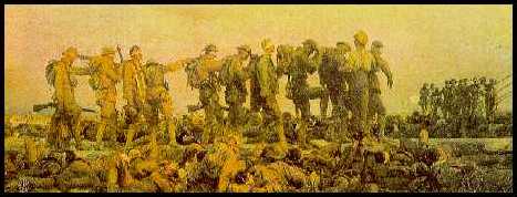
Sulfur mustard (History) was first synthesized in the early 1800s and was first used on the battlefield during World War I by Germany in July 1917. Despite its introduction late in that conflict, mustard produced the most chemical casualties, although fewer than 5% of the casualties who reached medical treatment facilities died. Italy allegedly used mustard in the 1930s against Abyssinia. Egypt apparently employed mustard in the 1960s against Yemen, and Iraq used mustard in the 1980s against Iran and the Kurds. Mustard is still considered a major threat agent of former Warsaw Pact countries and third world countries.
The United States manufactured mustard during World War I and World War II and maintains a stockpile that is currently undergoing destruction.
Nomenclature: Sulfur mustard manufactured by the Levinstein process contains up to 30% impurities (mostly sulfur) and is known as H. Mustard made by a distillation procedure is almost pure and is known as HD (distilled mustard). An early term for the German agent was HS (probably derived from the World War I slang term, Hun Stoffe).
PHYSICOCHEMICAL CHARACTERISTICS
Mustard is an oily liquid with a color ranging from light yellow to brown (description). Its odor is that of garlic, onion, or mustard (hence its name), but because of accommodation of the sense of smell, odor should not be relied on for detection. Under temperate conditions, mustard evaporates slowly and is primarily a liquid hazard, but its vapor hazard increases with increasing temperature. At 100oF or above, it is a definite vapor hazard. Mustard freezes at 57oF, and since a solid is difficult to disperse, mustard is often mixed with substances with a lower freezing point, e.g., Lewisite (the mixture is HL), or agent T, a closely related vesicant (the mixture is HT) so that the mixture will remain liquid at lower temperatures. The mixture HT also refers to mustard that has been thickened with small quantities of newer thickening agents.
The immediately-dangerous-to-life-and-health (IDLH) concentration of sulfur mustard (H) is 0.003 mg/m3. The M8A1 automatic chemical agent detector alarm is incapable of detecting mustard. However, liquid mustard turns M8 paper a ketchup red, and M9 paper will turn pink, red, reddish-brown, or purple when exposed to liquid nerve agents or vesicants, but does not specifically identify either the class of agent or the specific agent. The following detectors have the capacity to detect sulfur mustard (H) at the threshold limits given:
|
DETECTOR |
H |
|
M256A1 |
3.0 mg/min3 |
|
M272 (in water) |
2.0 mg/min3 |
|
MINICAMS |
0.00003 mg/min3 |
|
ICAD |
10.0 mg/min3 |
|
M18A2 |
0.5 mg/min3 |
|
M21 |
150 mg/min3 |
|
M90 |
0.2 mg/min3 |
|
M93A1 Fox |
0.01 - 1 mcg/l |
|
ACAMS |
0.003 mg/min3 |
|
Bubbler |
0.003 mg/min3 |
|
CAM |
0.1 mg/min3 |
|
DAAMS |
0.003 mg/min3 |
Because the odor of sulfur mustard may be faint or lost after accommodation, olfactory detection of the odor of mustard, garlic, onions, or horseradish is not a reliable indicator of mustard exposure. The activated charcoal in the canister of the chemical protective mask adsorbs mustard, as does the charcoal in the chemical protective overgarment. The butyl rubber in the chemical protective gloves and boots is impermeable to mustard. Proper wear of the chemical protective mask and the chemical protective ensemble affords full protection against sulfur mustard.
Mustard vapor and liquid readily penetrate thin layers of most fabrics (but not the chemical protective ensemble) to reach underlying skin. Although mustard dissolves relatively slowly in aqueous solutions such as sweat, the lipophilicity of mustard guarantees effective absorption through even intact skin. Penetration is rapid (1 to 4 mcg/cm2-min) and is enhanced by moisture, heat, and thin skin. This explains the otherwise baffling observation that World War I mustard burns involved the scrotum in 42% of cases, but the presumably more readily exposed hands in only 4% of cases. Ocular and respiratory routes of entry are also important, as is parenteral absorption in casualties with conventional wounds. Ingestion (enteral absorption) was an important route of entry for mustard in the sailors exposed outside Bari in World War II. Approximately 10% of the amount of mustard that begins to penetrate the skin will bind to the skin as "fixed" (reacted) mustard; the remaining 90% of the dose reaches the circulation and is systemically distributed as "free" (unreacted and hydrolyzed) mustard. Distribution to almost all organs and tissues including the kidneys, liver, intestines, and lungs occurs; although, because of dilutional effects and reactions of mustard in the bloodstream, clinical effects from systemic distribution are seen only at high doses. After intravenous administration, mustard disappears from the blood within seconds to minutes. Because of the rapid fixation of mustard to tissue, the fluid inside the blisters that eventually develop at the sites of skin contact contains no free mustard and does not pose a contamination hazard to health care providers. Mustard participates in a variety of biotransformative (metabolic) reactions in the body. Some of these reactions are catalyzed by enzymes, but most absorbed mustard reacts directly by forming covalent bonds (via alkylation) with DNA, RNA, proteins, components of cell membranes, and other macromolecules in the body. Mustard is eliminated primarily in the urine as by-products of alkylation.
The LCt50 of sulfur mustard dispersed as a vapor is 1500 mg-min/m3 in an unprotected group and 10,000 mg-min/m3 in a group with respiratory protection. This demonstrates not only the importance of respiratory protection, but also the fact that sufficient concentrations of vapor and sufficient exposure times render mustard vapor lethal, even in masked individuals. The LD50 of liquid mustard on the skin is 100 mg/kg. Thus, administration of 7 g (about a teaspoon) of liquid mustard to each member of a group of individuals weighing 70 kg would be expected to cause the death of half of those exposed. Although 7 g of a liquid applied evenly to the surface of the skin may cover approximately 20 to 25% of the total body surface area (BSA), the correlation between BSA involvement and deaths from mustard in the field is poor. One plausible reason for this discrepancy is that using BSA figures by themselves ignore the inhalational component of mustard exposure. Another conceivable explanation is that measurement solely of affected BSA neglects factors such as the thickness of coverage, subsequent spread, contact time, and continued exposure. A 10 mcg droplet of sulfur mustard can produce a small vesicle on exposed skin.
TOXICODYNAMICS
(MECHANISM OF ACTION)
Absorbed mustard must first dissolve in aqueous solution such as sweat or extracellular fluid. Although mustard molecules dissolve slowly in such solutions, once they dissolve they rapidly (within seconds to a minute or two) rearrange to form extremely reactive cyclic ethylene sulfonium ions that immediately bind to intracellular and extracellular enzymes, proteins, and other cellular components. Mustard has many biological actions, but the exact mechanism by which it produces tissue injury is not known. According to one prominent hypothesis, biological damage from mustard results from DNA alkylation and crosslinking in rapidly dividing cells, such as basal keratinocytes, mucosal epithelium, and bone marrow precursor cells. This leads to cellular death and inflammatory reaction, and, in the skin, protease digestion of anchoring filaments at the epidermal-dermal junction and the formation of blisters.
Mustard also possesses mild cholinergic activity, which may be responsible for effects such as early GI symptoms and miosis.
It should be re-emphasized that mustard reacts with tissue within minutes of entering the body and that blood, tissue, and blister fluid do not contain free mustard, nor do they represent a contamination risk for medical personnel.
Topical effects of mustard occur in the eye, airways, and skin. Systemically absorbed mustard may produce effects in the bone marrow, GI tract, and CNS. Direct injury to the GI tract may also occur following ingestion of the compound.
Combined data from United States forces in World War I and Iranians in the Iraq-Iran conflict suggest equal incidence of eye, airway, and skin involvement (between 80 and 90% for each). However, there were higher incidences of eye and lung damage in Iranian casualties than in World War I casualties, probably because of the larger amount of evaporation of the agent in the hot climate.
Skin: Erythema is the mildest and earliest form of skin injury after exposure to mustard (onset). It resembles sunburn and is associated with pruritus or burning, stinging pain. Erythema begins to appear in 2 to 48 hours after vapor exposure with time of onset dependent on Ct, ambient temperature and humidity, and skin site exposed. The skin sites most sensitive are the warm, moist locations with thinner skin such as the perineum, external genitalia, axillae, antecubital fossae, and neck.
Within the erythematous areas, small vesicles can develop which may later coalesce to form bullae. The typical bulla, or blister, is large, dome-shaped, thin-walled, translucent, yellowish, and surrounded by erythema. The blister fluid is clear, at first thin and straw-colored, but later yellowish and tending to coagulate. The fluid does not contain mustard and is not a vesicant.
At extremely high doses such as those from liquid exposure, lesions may develop a central zone of coagulation necrosis with blister formation at the periphery. These lesions take longer to heal and are more prone to secondary infection than the uncomplicated lesions seen at lower exposure levels.
Pulmonary: The primary airway lesion from mustard is necrosis of the mucosa with later damage to the musculature of the airways if the amount of agent is large. The damage begins in the upper airways and descends to the lower airways in a dose-dependent manner. Usually the terminal airways and alveoli are affected only as a terminal event. Pulmonary edema is not usually present unless the damage is very severe, and then it usually is hemorrhagic.
The earliest effects from mustard, perhaps the only effects from a low Ct, involve the nose, sinuses, and pharynx. There may be irritation or burning of the nares, epistaxis, sinus pain or irritation, and irritation or soreness of the pharynx. As the Ct increases, other effects occur - laryngitis with voice changes and a nonproductive cough. Damage to the trachea and upper bronchi leads to a cough productive of sputum. Lower airway involvement causes dyspnea and an increasingly severe cough with increased quantities of sputum. Terminally, there may be necrosis of the smaller airways with hemorrhagic edema into surrounding alveoli. This hemorrhagic pulmonary edema is rarely a feature.
Necrosis of the airway mucosa with resulting inflammation can cause pseudomembrane formation. Pseudomembranes may occur from the most proximal parts of the airways to the most distal portions. These membranes may cause local airway obstruction at the sites of formation, and detachment may lead to obstruction of lower airways. (Airways)
The cause of death in mustard poisoning is commonly respiratory failure. Mechanical obstruction by pseudomembranes and agent-induced laryngospasm are important causes of death in the first 24 hours after exposure. Deaths occurring from the third to the sixth day after exposure result from secondary bacterial pneumonia caused by bacterial invasion of denuded respiratory mucosa and necrotic debris. Agent-induced bone marrow suppression is a contributory factor in later, septic deaths from pneumonia.
Eyes: The eyes are the organs most sensitive to mustard vapor injury. The latent period is shorter for eye injury than for skin injury and is also Ct dependent.
After low-dose vapor exposure, irritation evidenced by reddening of the eyes may be the only effect. As the dose increases, the spectrum of injury includes progressively more severe conjunctivitis, photophobia, blepharospasm, pain, and corneal damage.
Blisters do not normally form in the eyes. Instead, swelling and loosening of corneal epithelial cells lead to corneal edema and clouding with leukocytes (which affects vision). Corneal vascularization with secondary edema may last for weeks. Scarring between the iris and lens may follow severe effects; this scarring may restrict pupillary movements and may predispose victims to glaucoma.
The most severe damage is caused by liquid mustard from airborne droplets or by self-contamination. After extensive eye exposure, severe corneal damage with possible perforation of the cornea and loss of the eye can occur. Eye loss also results from panophthalmitis if appropriate therapy is not instituted.
During World War I, mild conjunctivitis accounted for 75% of eye injuries, with recovery in one to two weeks. Moderate conjunctivitis with minimal corneal involvement, blepharospasm, edema of the lids and conjunctivae, and orange-peel roughening of the cornea accounted for 15% of the cases, with recovery in four to six weeks. Severe corneal involvement accounted for 10% of the cases. Those with permanent corneal damage accounted for less than 1% of cases. About 0.1% of these severe casualties would meet the criteria for legal blindness today.
Miosis noted after mustard exposure in both humans and experimental animals is probably from the cholinomimetic activity of mustard.
Gastrointestinal (GI) tract: The mucosa of the GI tract is very susceptible to mustard damage, either from systemic absorption or ingestion of the agent. However, reports of severe GI effects from mustard poisoning are relatively infrequent.
Mustard exposure, even exposure to a small amount, will often cause nausea, with or without vomiting, lasting 24 hours or less. The nausea and vomiting appear not to be a direct effect of the agent on the GI tract, but rather they are from a stress reaction, a nonspecific reaction to the odor, or cholinergic stimulation by mustard. Further GI symptoms are usually minimal unless the exposure was severe (even then, GI signs are not common) or exposure resulted from ingestion of contaminated food or drink. Diarrhea has been reported; constipation is equally common. Diarrhea (rarely bloody) and vomiting beginning days after a high-dose exposure imply a poor prognosis
Central nervous system (CNS): The CNS effects of mustard remain poorly defined. Animal work demonstrated that mustards (particularly the nitrogen mustards) are convulsants, and there are several human case reports describing victims who were exposed to very large amounts and had neurological effects within several hours after exposure just prior to death. Reports from World War I, and again from Iran, described people exposed to small amounts of mustard who appeared sluggish, apathetic, and lethargic. These reports suggest that minor psychological problems could linger for a year or longer.
Mustard binds irreversibly to tissue within several minutes after contact (onset). If decontamination is not done immediately after exposure, there is no way to prevent injury, although later decontamination might prevent a more severe lesion.
The clinical effects of mustard are delayed. Signs and symptoms may appear as early as 2 hours after a high-dose exposure, whereas following a low-dose vapor exposure, the latent or asymptomatic period may extend to 48 hours. There are several reports of individuals exposed to very large amounts who died within hours; this type of occurrence is extremely rare. The typical onset time is between four and eight hours. The concentration (C) of the mustard vapor, time (t) of exposure, ambient weather, and body site exposed are factors in the onset time.
It must be emphasized that mustard causes tissue damage within several minutes after contact without causing any concomitant clinical effects, e.g., burning or erythema. Because of the lack of immediate effects, the contaminated person is often unaware of the exposure and does not decontaminate. To prevent injury, decontamination must be done immediately after contact. Later decontamination may prevent further damage, absorption, or spread of the agent.
Table: Effects of Mustard Vapor
|
ORGAN |
SEVERITY |
EFFECTS |
ONSET OF FIRST EFFECT |
|
Eye |
Mild |
Tearing, itchy, burning, gritty feeling |
4-12 hours |
|
|
Moderate |
Above, plus reddening, swelling of lids, moderate pain |
3-6 hours |
|
|
Severe |
Marked swelling of lids, possible cornea damage, severe pain |
1-2 hours |
|
Airways |
Mild |
Runny nose, sneezing, nosebleed, hoarseness, hacking cough |
12-24 hours |
|
|
Severe |
Above, plus severe productive cough, shortness of breath |
2-4 hours |
|
Skin |
Mild to Severe |
Erythema (redness), blisters |
2-24 hours |
Of the three vesicant agents, mustard is the only one that does not cause immediate pain. The casualty is asymptomatic until the lesion becomes apparent hours later.
Lewisite and phosgene oxime, in contrast, cause immediate pain or irritation to the eye, skin, or respiratory tract. This causes sufficient stimulus to decontaminate immediately or to mask.
Isolated small blisters or a small group of blisters suggest possible exposure to mustard, to plants such as poison ivy or poison oak, drugs, or other substances. The physical characteristics of the lesion are not distinctive; therefore, the history of exposure is invaluable.
Although the blisters of mustard and Lewisite are slightly different (there is less erythema around the Lewisite blister), this information is of little value in individual cases. (Differential Diagnosis) (Diagnostics)
Leukocytosis occurs during the first day, and the magnitude of increase in leukocytes during the subsequent days correlates roughly with the amount of tissue injury, primarily to skin or pulmonary tissue. If systemic absorption is large, leukocytes in the peripheral blood will decrease beginning on day three to day five; this decrease indicates damage to precursor cells in the blood-forming organs. The fall may be precipitate, e.g., a decrease of 5000 to 10,000 cells/day. If the marrow damage is severe, erythrocytes and thrombocytes may later decrease, but the casualty usually recovers or dies before this is apparent. A leukocyte count of 500 or fewer is a sign of an unfavorable prognosis.
Signs of a chemical pneumonitis may appear within the first two to three days after inhalation exposure. Leukocytosis, fever, and sputum production suggest a bacterial process, but within this time period sputum cultures are usually negative for pathogens. Organisms commonly invade the damaged airway tissue at days three to five. A change in the fever pattern, an increase in leukocytosis, and a change in the character of the sputum in this time period suggest a bacterial process. Sputum Gram Stain and culture should be done for identification of the specific organism.
Damaged skin should be cultured routinely, particularly if there is an increase in the exudate or in the inflammatory reaction.
Although GI bleeding is unusual, declining hematocrit values should prompt serial analyses of stool for occult blood.
Thiodiglycol, a urinary metabolite of sulfur mustard, can be measured by the Theater Army Medical Laboratory (TAML), which will be deployed. There is no clinical laboratory test for mustard in blood or tissue, nor is one expected, as mustard is biotransformed and bound to tissues within minutes after absorption. However, ways to measure blood and tissue adducts produced in the body after reaction with sulfur mustard are being studied.
The management of a patient exposed to mustard may be simple, as in the provision of symptomatic care for a sunburn-like erythema, or extremely complex, as providing total management for a severely ill patient with burns, immunosuppression, and multi-system involvement. Suggested therapeutic measures for each organ system are provided below. Guidelines for general patient care are not intended to take the place of sound clinical judgment, especially in the management of complicated cases.
Skin: Erythema should be treated with calamine or other soothing lotion or cream (e.g., 0.25% camphor and menthol, calamine) to reduce burning and itching. Small blisters (under 1-2 cm) should be left intact, but because larger ones will eventually break (the blister fluid does not contain mustard), they should be carefully unroofed. Denuded areas should be irrigated three to four times daily with saline, another sterile solution, or soapy water and then liberally covered with a topical antibiotic such as silver sulfadiazine or mafenide acetate to a thickness of 1-2 mm. If an antibiotic cream is not available, sterile petrolatum will be useful. Modified Dakins solution (sodium hypochlorite) was used in World War I and in Iranian casualties for irrigation and as an antiseptic.
Multiple or large areas of vesication suggest the need for hospitalization and whirlpool bath irrigation.
Systemic analgesics should be used liberally, particularly before manipulation of the patient or irrigation of the burn areas. Systemic antipruritics such as trimeprazine should be tried if needed. Monitoring of fluids and electrolytes is important in any sick patient, but it must be recognized that fluid loss is not of the magnitude seen with thermal burns. Clinicians accustomed to treating patients with thermal burns must resist the temptation to overhydrate a mustard casualty with a similar amount of burned body surface.
Eyes: Conjunctival irritation from a low Ct will respond to any of a number of available ophthalmic solutions after the eyes are thoroughly irrigated. Regular application of homatropine (or other anticholinergic drug) ophthalmic ointment will reduce or prevent future synechiae formation. A topical antibiotic applied several times a day will reduce the incidence and severity of infection. Vaseline or a similar substance should be applied to the edges of the lids regularly to prevent them from sticking together. This prevents adhesions and later scarring during healing and also permits drainage of any underlying infection or pus. Topical analgesics may be useful initially if blepharospasm is too severe to permit an adequate examination, but topical analgesics should otherwise be avoided and systemic analgesics should be given for eye pain. Topical steroids are not of proven value, but their use during the first day or two might reduce inflammation. Further use should be relegated to an ophthalmologist. Sunglasses may reduce discomfort from photophobia
The patient should be constantly reassured that complete healing and restoration of vision will be the outcome.
Pulmonary: Upper airway symptoms (sore throat, nonproductive cough, and hoarseness) may respond to steam inhalation and cough suppressants. Although a productive cough and dyspnea accompanied by fever and leukocytosis occurring 12 to 24 hours after exposure may suggest a bacterial process to the clinician, he must resist the urge to use antibiotics for this process, which in fact is a sterile bronchitis or pneumonitis. Infection often occurs on about the third day. Its presence is signaled by an increased fever, an increase in the pulmonary infiltrate by x-ray, and an increase in sputum production and a change in sputum character to purulent. Appropriate antibiotic therapy should await confirmation of the clinical impression by positive sputum studies (Gram stain and culture)
Intubation should be performed early before laryngeal spasm or edema make it difficult or impossible. Intubation permits better ventilation and facilitates suction of the necrotic and inflammatory debris. Oxygen may be needed, and early use of PEEP or CPAP may be of benefit. If there is a suggestion of pseudomembrane formation, bronchoscopy should be done to permit suctioning of the necrotic debris by direct vision.
Bronchodilators may be of benefit for bronchospasm. If they fail, steroids may be tried. There is little evidence that the routine use of steroids is beneficial. The need for continuous use of assisted or controlled ventilation suggests a poor prognosis.
Death often occurs between the fifth and tenth day after exposure because of pulmonary insufficiency and infection complicated by a compromised immune response from agent-induced bone marrow damage.
Gastrointestinal: Atropine (0.4-0.6 mg, i.m. or i.v.), another anticholinergic drug or antiemetic, should control the early nausea and vomiting. Prolonged vomiting or voluminous diarrhea beginning days after exposure suggests direct involvement of the GI tract by severe systemic poisoning, a poor prognostic sign.
Bone Marrow: Sterilization of the gut by nonabsorbable antibiotics should be considered to reduce the possibility of sepsis from enteric organisms. Cellular replacement (bone marrow transplants or transfusions) may be successful, as intact mustard does not persist beyond the few minutes following absorption and would not damage the new cells.
General: A patient severely ill from mustard poisoning requires the general supportive care provided for any severely ill patient, as well as the specific care given to a burn patient. Liberal use of systemic analgesics and antipruritics, as needed, maintenance of fluid and electrolyte balance, and other supportive measures are necessary. Parenteral food supplements including vitamins may also be helpful.
Other: Sulfur donors such as sodium thiosulfate decreased systemic effects and elevated the LD50 when given before exposure or within 20 minutes after exposure in experimental animals. Activated charcoal given orally to casualties was of no value. Hemodialysis was not only ineffective, but was actually harmful in several casualties. The rapid biotransformation of the mustard molecule suggests that none of these measures would be beneficial hours or days after exposure.
Most mustard casualties will be triaged as delayed. Those with skin lesions covering several percent to 50% of the BSA will require further medical care but do not need immediate life-saving assistance. (In contrast, patients with thermal burns covering 20 to 70% of their BSA are considered immediate because of their fluid requirements.) Those with mild to moderate pulmonary effects will also eventually require further care, but are not in the immediate category for triage. Eye injuries from other causes require immediate care, but by the time the mustard eye lesion develops, there is no possibility of reducing the injury. These casualties are also in the delayed category.
Patients with skin lesions covering a small percent of BSA (under 5%), when the lesions are not in vital areas (a burn on the face might prevent mask donning), are triaged as minimal. Clinical judgement should dictate whether these patients should be evacuated for care or whether they can return to duty. The tactical situation will also be a factor in the decision. Patients with minor eye injuries to include irritation and reddening can be treated and returned to duty. Those with slight upper respiratory complaints of a hacking cough and an irritated throat which developed 12 hours or longer after exposure might be given symptomatic therapy and returned to duty.
The only mustard casualties who might be triaged as immediate are those with moderately severe to severe pulmonary signs and symptoms. Two factors should temper this decision. (1) Casualties who develop severe pulmonary effects within four to six hours of exposure will probably not survive despite maximal medical care, and it might be better to expend limited medical resources elsewhere. (2) If evacuation to a maximal medical care facility is required, the casualty may survive the lengthy trip, but during the delay his lesion may progress to an irreversible stage.
A mustard casualty who has severe pulmonary effects that developed within four to six hours of exposure should be triaged as expectant. A casualty who has over 50% BSA burns from mustard liquid might also be categorized as expectant, but this decision would depend on available medical resources at the far rear echelons of medical care. (The LD50 for liquid mustard is about 7 grams, or between one and one and a half teaspoons of liquid. This amount will cover about 25% BSA, so an individual with a 50% BSA burn could possibly have two LD50s on his skin. This person might be saved, but at great expenditure of medical resources.)
Casualties with minor skin, eye, or pulmonary injuries might be returned to duty as soon as they are given symptomatic therapy at a medical facility. The range of return to duty times for those with more severe but treatable injuries is from one week to a year or longer.
Those with eye injuries should recover in one to three weeks, except for the low percentage of casualties with severe injuries or complications. Casualties with mild to moderate pulmonary injuries should return in a week to a month. Healing of mild skin lesions will enable the casualty to return within several weeks, but patients with large skin lesions will require hospitalization for many months.
Repeated symptomatic exposures to mustard over a period of years (as in manufacturing workers) seem to be well established as a causal factor in an increased incidence of upper airway cancer. However, the association between a single exposure to mustard and airway cancer is not well established. A single, severe exposure to mustard may have contributed to other airway problems, such as chronic bronchitis, based on World War I data. A new complication seen in Iranian casualties from the Iran-Iraq War in the 1980s was late-onset tracheobronchial stenosis, which presumably would have been seen in World War I casualties had antibiotic therapy been available to allow those who died from secondary bacterial pneumonia to survive.
Several eye diseases, such as chronic conjunctivitis and delayed keratitis, may follow a single, severe exposure of the eye to mustard. Skin scarring and pigment changes may follow a severe skin lesion from mustard; cancer sometimes develops in scarred skin.
Mustard is classed as a mutagen and carcinogen based on laboratory studies. However, there are no data to implicate mustard as a reproductive toxin in man, and there is no evidence that mustard is a causative factor in nonairway, non-skin cancer in man.
L
Signs and Symptoms: Lewisite causes immediate pain or irritation of skin and mucous membranes. Erythema and blisters on the skin and eye and airway damage similar to those seen after mustard exposures develop later.
Detection: M256A1, M272 water testing kit, MINICAMS, the ICAD, M18A2, M21 remote sensing alarm, M90, M93A1 Fox, Bubbler, CAM, and DAAMS (but NOT the M8A1 automatic chemical agent alarm), M8 paper, or M9 paper.
Decontamination: M291, 0.5% hypochlorite, water in large amounts.
Management: immediate decontamination; symptomatic management of lesions the same as for mustard lesions; a specific antidote (BAL) will decrease systemic effects.
Lewisite is a vesicant that damages the eyes, skin, and airways by direct contact. After absorption, it causes an increase in capillary permeability to produce hypovolemia, shock, and organ damage. Exposure to Lewisite causes immediate pain or irritation, although lesions require hours to become full-blown. Management of a Lewisite casualty is similar to management of a mustard casualty, although a specific antidote, British-Anti-Lewisite (BAL, dimercaprol), will alleviate some effects.
Dr. Wilford Lee Lewis first synthesized Lewisite in 1918, but production was too late for its use in World War I. It has not been used in warfare, although some countries may stockpile it. Lewisite is sometimes mixed with mustard to achieve a lower freezing point of the mixture for ground dispersal and aerial spraying.
PHYSICOCHEMICAL CHARACTERISTICS
Lewisite is an oily, colorless liquid with the odor of geraniums. It is more volatile than mustard.
The immediately-dangerous-to-life-and-health (IDLH) concentration of Lewisite (L) is 0.003 mg/m3. The M8A1 automatic chemical-agent detector alarm is incapable of detecting Lewisite. However, liquid Lewisite turns M8 paper a ketchup red, and M9 paper will turn pink, red, reddish-brown, or purple when exposed to liquid nerve agents or vesicants, but does not specifically identify either the class of agent or the specific agent. The following detectors have the capacity to detect Lewisite (L) at the threshold limits given:
|
DETECTOR |
L |
|
M256A1 |
14.0 mg/min3 |
|
M272 (in water) |
2.0 mg/min3 |
|
MINICAMS |
0.0006 mg/min3 |
|
ICAD |
10.0 mg/min3 |
|
M18A2 |
10.0 mg/min3 |
|
M21 |
150.0 mg/min3 |
|
M90 |
0.2 mg/min3 |
|
M93A1 Fox |
10 - 100 mcg/l |
|
Bubbler |
0.003 mg/min3 |
|
CAM |
2.0 mg/min3 |
|
DAAMS |
0.003 mg/min3 |
Because the odor of Lewisite may be faint or lost after accommodation, olfactory detection of the odor of geraniums is not a reliable indicator of Lewisite exposure. The activated charcoal in the canister of the chemical protective mask adsorbs Lewisite, as does the charcoal in the chemical protective overgarment. Lewisite attacks the butyl rubber in the chemical protective gloves and boots, which nevertheless are expected to protect against field concentrations of Lewisite until they can be exchanged for fresh gloves and boots. Proper wear of the chemical protective mask and chemical protective ensemble affords full protection against Lewisite.
Lewisite is readily absorbed from the skin, eyes, and respiratory tract, as well as by ingestion and via wounds. It is systemically distributed to almost all organs and tissues of the body where it participates in a variety of chemical reactions. It is eventually eliminated primarily as reaction products in the urine.
TOXICITY
Lewisite causes nasal irritation at a Ct of about 8 mg-min/m3, and its odor is noted at a Ct of about 20 mg-min/m3. Lewisite causes vesication and death from inhalation at the same Ct as mustard. Liquid Lewisite causes vesication at about 14 mcg, and the LD50 of liquid Lewisite applied to the skin is about 2.8 grams.
TOXICODYNAMICS (MECHANISM OF ACTION)
Although Lewisite contains trivalent arsenic and combines with thiol groups in many enzymes, its exact mechanism of biological activity is unknown.
Organ Systems: Unlike mustard, Lewisite vapor or liquid causes immediate pain or irritation. A person with a droplet of Lewisite on his skin will note the burning and will immediately take steps to try and remove it. The vapor is so irritating that a person will seek to mask or leave the contaminated area if possible. Because this warning causes the person exposed to take immediate steps to decontaminate, the Lewisite lesion will probably not be as severe as the lesion from mustard, as exposure to mustard is often undetected and decontamination is not done.
There are almost no data on humans exposed to Lewisite. The following information is based on animal investigations.
Skin: Within about five minutes after contact, liquid Lewisite will produce a grayish area of dead epithelium. Erythema and blister formation follow more rapidly than in a similar lesion from mustard, although the full lesion does not develop for 12 to 18 hours. The lesion has more tissue necrosis and tissue sloughing than does a mustard lesion.
Eyes: Lewisite causes pain and blepharospasm on contact. Edema of the conjunctiva and lids follows, and the eyes may be swollen shut within an hour. Iritis and corneal damage may follow if the dose is high. Liquid Lewisite causes severe eye damage within minutes of contact.
Respiratory: The extreme irritancy of Lewisite to the nasal area and upper airways causes the person to mask or exit the area. Scanty data indicate that Lewisite causes the same airway signs and symptoms as does mustard. The airway mucosa is the primary target, and damage progresses down the airways in a dose-dependent manner. Pseudomembrane formation is prominent. Pulmonary edema, which occurs rarely and usually only to a minimal degree after mustard exposure, may complicate exposure to Lewisite.
Other: Available data suggest that Lewisite causes an increase in permeability of systemic capillaries with resulting intravascular fluid loss, hypovolemia, shock, and organ congestion. This may lead to hepatic or renal necrosis with more prominent GI effects (including vomiting and diarrhea) than after mustard.
Physical Findings: The findings are similar to those caused by mustard. As noted, the tissue damage at the site of the skin lesion may be more severe.
Pain and irritation from either liquid or vapor Lewisite are immediate. Early tissue destruction is more obvious than after mustard, but the lesion is not full-blown for 12 hours or longer.
Although differences have been reported between the skin lesions from mustard and Lewisite (less surrounding erythema and more tissue destruction characterize Lewisite blisters), these are of little diagnostic assistance in a single patient. The history of immediate pain on contact is absent after mustard exposure and present after Lewisite or phosgene oxime exposures.
Other substances cause erythema and blisters, and often the history of exposure is the most helpful tool in diagnosis.
There is no specific diagnostic test for Lewisite. Leukocytosis, fever, and other signs of tissue destruction will occur.
Early decontamination is the only way of preventing or lessening Lewisite damage. Since this must be accomplished within minutes after exposure, this is self-aid rather than medical management.
The guidelines for the management of a mustard casualty will be useful. Lewisite does not cause damage to hematopoietic organs as mustard does; however, fluid loss from the capillaries necessitates careful attention to fluid balance.
British-Anti-Lewisite (BAL, dimercaprol) was developed as an antidote for Lewisite and is used in medicine as a chelating agent for heavy metals. There is evidence that BAL in oil, given intramuscularly, will reduce the systemic effects of Lewisite. However, BAL itself causes some toxicity, and the user should read the package insert carefully. British-Anti-Lewisite skin and ophthalmic ointment decreases the severity of skin and eye lesions when applied immediately after early decontamination; however, neither is currently manufactured.
Casualties should be triaged using the guidelines for triage of mustard patients.
Casualties with minor skin lesions who receive symptomatic therapy can be returned to duty quickly. Because Lewisite generally causes more tissue damage than mustard, casualties with eye and larger skin lesions should be triaged as delayed and evacuated. Whether to triage those with pulmonary injury as immediate, delayed, or expectant depends on the severity of the injury and how quickly after exposure it occurred.
PHOSGENE OXIME
CX
Signs and Symptoms: immediate burning and irritation followed by wheal-like skin lesions and eye and airway damage.
Detection: M256A1, M18A2, M90, and M93 Fox (but NOT the M272 water testing kit), MINICAMS, the ICAD, M21 remote sensing alarm, Bubbler, CAM, DAAMS, the M8A1 automatic chemical agent alarm, M8 paper, or M9 paper.
Decontamination: water in large amounts, 0.5% hypochlorite, M291.
Management: immediate decontamination, symptomatic management of lesions.
Phosgene oxime (CX) is an urticant or nettle agent that causes a corrosive type of skin and tissue lesion. It is not a true vesicant since it does not cause blisters. The vapor is extremely irritating, and both the vapor and liquid cause almost immediate tissue damage upon contact. There is very scanty information available on CX.
There is no current assessment of the potential of CX as a military threat agent.
PHYSICOCHEMICAL CHARACTERISTICS
Phosgene oxime is a solid at temperatures below 95oF, but the vapor pressure of the solid is high enough to produce symptoms. Traces of many metals cause it to decompose; however, it corrodes most metals.
The immediately-dangerous-to-life-and-health (IDLH) concentration of CX has not been defined. The M272 water testing kit, MINICAMS, ICAD, M21 remote sensing alarm, CAM, ACAMS, DAAMS, and M8A1 automatic chemical-agent detector alarm are incapable of detecting CX. Likewise, M8 and M9 paper should not be depended upon to detect this agent. The M256A1 detector ticket reacts to the presence of CX, but the detection threshold is not known with certainty. The following detectors have the capacity to detect CX at the threshold limits given:
|
DETECTOR |
CX |
|
M18A2 |
0.5 mg/min3 |
|
M90 |
0.15 mg/min3 |
|
M93A1 Fox |
10 - 100 mcg/l |
Because the odor of phosgene may be faint or lost after accommodation, olfactory detection of a pepperish or pungent odor is not a reliable indicator of the presence of CX. The activated charcoal in the canister of the chemical protective mask adsorbs Lewisite, as does the charcoal in the chemical protective overgarment. Phosgene oxime may attack the butyl rubber in the chemical protective gloves and boots, which nevertheless are expected to protect against field concentrations of CX until they can be exchanged for fresh gloves and boots. Proper wear of the chemical protective mask and chemical protective ensemble affords full protection against CX.
The toxicokinetics of CX are not known in detail. Penetration of exposed surfaces is rapid, and systemic distribution to most organs and tissues, including the GI tract, is probably important.
TOXICITY
The estimated LCt50 by inhalation is 1500-2000 mg·min/m3. The LD50 for skin exposure has been estimated as 25 mg/kg.
TOXICODYNAMICS (MECHANISM OF ACTION)
The mechanism by which CX causes biological effects is unknown. (Clinical Effects)
Skin: Phosgene oxime liquid or vapor causes pain on contact, which is followed in turn by blanching with an erythematous ring in 30 seconds, a wheal in 30 minutes, and necrosis later. The extreme pain may persist for days.
Eyes: Phosgene oxime is extremely painful to the eyes. The damage is probably similar to that caused by Lewisite.
Pulmonary: Phosgene oxime is very irritating to the upper airways. This agent causes pulmonary edema after inhalation and after skin application.
Other: Some animal data suggest that CX may cause hemorrhagic inflammatory changes in the GI tract.
Phosgene oxime causes immediate pain and irritation to all exposed skin and mucous membranes. The time course of damage to other tissue probably parallels that of damage to the skin.
Other causes of urticaria and skin necrosis must be considered. Common urticants do not cause the extreme pain that CX does.
There are no distinctive laboratory findings.
Management is supportive. The skin lesion should be managed in the same way that a necrotic ulcerated lesion from another cause would be managed.
Because of the continuing pain, most casualties should be placed in the delayed category and evacuated.
The decision to return a CX casualty to duty should be based on healing of the lesion(s) and the casualty's freedom from discomfort.